Anatomy and Physiology of ABC
Understanding basic anatomy and physiology is essential for effective first aid. This knowledge helps you understand what's happening in the body during emergencies and why certain first aid techniques work.
The ABC approach is the foundation of emergency assessment and care. Each component represents a critical body system that must function properly to sustain life.
## Why ABC Prioritization Matters
The ABC approach reflects the body's survival priorities:
- Airway - Without an open airway, oxygen cannot enter
- Breathing - Without breathing, oxygen cannot reach the blood
- Circulation - Without circulation, oxygenated blood cannot reach vital organs
## Airway
The airway is the pathway that allows air to travel from the outside environment to the lungs and back out again. It's the first priority in emergency care because without a clear airway, breathing cannot occur.
Anatomy of the Airway
Upper Airway Components:
- Nose and Mouth - Primary entry points for air
- Pharynx (Throat) - Shared pathway for breathing and swallowing
- Larynx (Voice Box) - Contains vocal cords and entrance to trachea
- Epiglottis - acts as a protective flap during swallowing
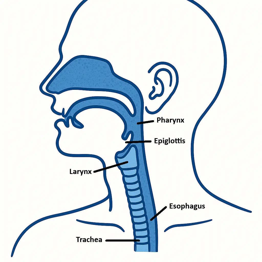
Upper Airway Anatomy
Lower Airway Components:
- Trachea (Windpipe) - Main tube leading to the lungs
- Bronchi - Two main branches (left and right)
- Bronchioles - Smaller branching tubes within the lungs
- Alveoli - Tiny air sacs where gas exchange occurs
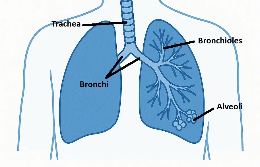
Lower Airway Anatomy
First Aid Assessment and Management
Airway Assessment:
- Look - Check for visible obstructions, chest movement
- Listen - Normal breathing sounds vs. snoring, wheezing, or silence
- Feel - Air movement from nose/mouth
Opening the Airway:
- Head-tilt, chin-lift - For unresponsive patients (no suspected spinal injury)
- Jaw-thrust - When spinal injury is suspected
## Breathing
Breathing is the mechanical process of moving air in and out of the lungs, combined with the vital gas exchange that occurs at the cellular level. Effective breathing requires both an open airway and proper lung function.
Inside the Lungs
Oxygen enters the blood: Inhaled air in the alveoli has a higher concentration of oxygen than the blood arriving from the body. Because of this concentration gradient, oxygen diffuses across the thin walls of the alveoli and capillaries into the bloodstream.
Carbon dioxide leaves the blood: The blood arriving at the lungs is rich in carbon dioxide, a waste product from the body's tissues. The concentration of carbon dioxide is higher in the blood than in the alveolar air. Therefore, carbon dioxide diffuses from the blood into the alveoli to be carried out of the body when you exhale.
At the body's tissues
Oxygen leaves the blood: The oxygenated blood then travels to the rest of the body. At the body's tissues, the concentration of oxygen is higher in the blood than in the cells. Oxygen diffuses from the blood into the cells to be used for cellular respiration.
Carbon dioxide enters the blood: As cells perform their functions, they produce carbon dioxide, creating a higher concentration of it in the cells than in the blood. Carbon dioxide then diffuses from the cells into the bloodstream to be transported back to the lungs.
Breathing Mechanics
Inspiration (Inhalation)
- Diaphragm contracts and moves downward
- Rib cage expands outward
- Lung volume increases, creating negative pressure
- Air rushes into lungs
Expiration (Exhalation)
- Diaphragm relaxes and moves upward
- Rib cage contracts inward
- Lung volume decreases, creating positive pressure
- Air is pushed out of lungs
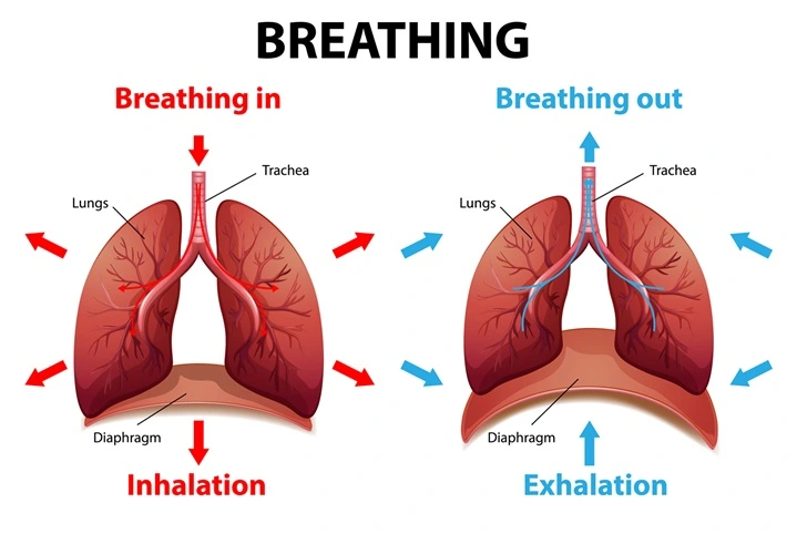
Breathing Mechanics (Designed by Freepik)
Air Composition: Inhaled vs. Exhaled
Understanding the difference between the air we breathe in and the air we breathe out helps explain why rescue breathing can be effective in first aid situations.
Inhaled Air (Atmospheric Air):
- Oxygen (O₂): ~21%
- Nitrogen: 78%
- Carbon Dioxide (CO₂) and Other gases: ~1%
Exhaled Air:
- Oxygen (O₂): ~16-17%
- Nitrogen: 78%
- Carbon Dioxide (CO₂): ~4-5%
- Other gases: Remainder
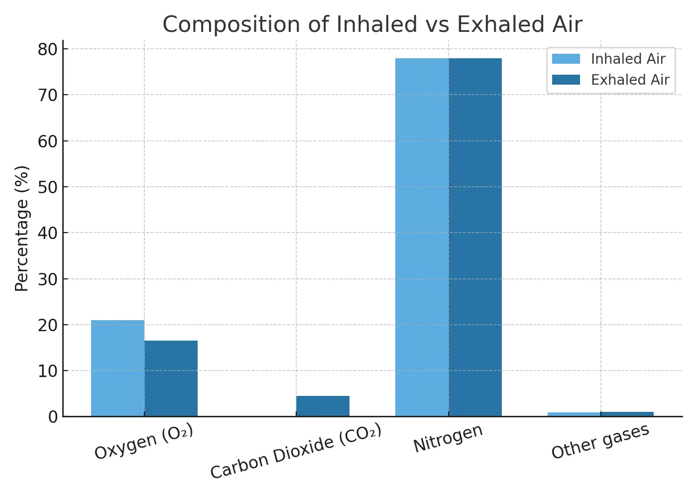
Inhaled and Exhaled Air Composition
Key Points for First Aid:
- Exhaled air still contains 16-17% oxygen - enough to sustain life during rescue breathing
- Normal air contains 21% oxygen - the body only uses about 4-5% during breathing
- This oxygen difference makes rescue breathing effective
How CPR Works
CPR is a combination of rescue breath and compression. Rescue breath is blowing air through the airway to the lungs as the oxygen source. The compression on the chest is helping to pump the blood in which help bringing the oxygenated blood from the lungs to the whole body.
Normal vs. Abnormal Breathing
Normal Breathing Characteristics:
- Rate: 12-20 breaths per minute (adults)
- Rhythm: Regular and consistent intervals
- Depth: Adequate chest rise and fall
- Effort: Effortless and quiet
- Pattern: Smooth, coordinated movement
## Circulation
Circulation is the continuous movement of blood through the cardiovascular system, delivering oxygen and nutrients to all body tissues while removing waste products. It's critical for survival.
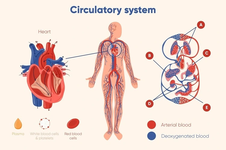
Circulatory System (Designed by Freepik)
The Heart:
- Size: Approximately the size of a closed fist
- Location: Center of chest, slightly left of midline
- Structure: Four-chambered muscular pump
Blood Vessels:
- Arteries - Carry oxygenated blood away from heart (except pulmonary artery)
- Veins - Return deoxygenated blood to heart (except pulmonary veins)
- Capillaries - Microscopic vessels where gas and nutrient exchange occurs
Physiology of Circulation
Think of your circulatory system as a delivery service that never stops working. Here's how it works in simple terms:
How Circulation Works - Step by Step:
1. The Heart is Your Body's Pump
- Your heart beats about 100,000 times per day
- Each beat pushes blood through your body
- It works like a two-sided pump with four chambers
2. The Journey Begins
- Step 1: The left side of your heart pumps fresh, oxygen-rich blood out through arteries
- Step 2: This bright red blood travels through smaller and smaller tubes until it reaches tiny capillaries
- Step 3: At the capillaries, the blood drops off oxygen and nutrients to your body's cells
- Step 4: The blood picks up waste products (like carbon dioxide) from the cells
3. The Return Trip
- Step 5: Now the blood is dark red because it's carrying waste instead of oxygen
- Step 6: This "used" blood travels back through veins to the right side of your heart
- Step 7: The right side of your heart pumps this blood to your lungs
- Step 8: In the lungs, the blood drops off carbon dioxide and picks up fresh oxygen
4. The Cycle Repeats
- The freshly oxygenated blood returns to the left side of your heart
- The cycle starts all over again
- This happens continuously, 24/7, for your entire life
Why This Matters for First Aid:
- If the heart stops → Blood stops moving → Cells don't get oxygen → Brain cells start dying in 4-6 minutes
- If someone is bleeding heavily → Less blood available to carry oxygen → Body goes into shock
- This is why CPR works → Chest compressions manually pump the heart to keep blood moving
Critical Timing: Brain Cell Death
Brain cells begin to die after 4-6 minutes without oxygen. After 10 minutes without oxygen, irreversible brain damage is likely to occur. This narrow window emphasizes the importance of immediate CPR and rapid emergency response.
First Aid Assessment and Management
Signs of Good Circulation:
- Pulse: Strong, regular, appropriate rate
- Skin color: Pink or normal skin tone
- Skin temperature: Warm to touch
- Mental status: Alert and oriented
- Blood pressure: Within normal range
Signs of Poor Circulation:
- Pulse: Weak, rapid, irregular, or absent
- Skin color: Pale, blue (cyanotic), or gray
- Skin temperature: Cool, cold, or clammy
- Mental status: Confused, anxious, or unconscious
- Blood pressure: Low or undetectable
Pulse Assessment Locations:
- Radial pulse - Wrist, thumb side
- Carotid pulse - Neck, beside Adam's apple
- Brachial pulse - Upper arm (infants)